
| Material type | Diameters available, µm |
| SiO2 | 2.01; 3.5; 4.63; 6.62; 10; 15; 28.75 |
| PMMA | 1.5; 3.36; 6.44; 10; 108 |
| BSG | 2; 5; 10; 20 |
| Gold | 1.5 - 3; 3 - 5.5; 5.5 - 9 |
| Glass beads | 10 - 50 |
| Barium Titanate Solid Glass | 74 - 90; 90 - 106 |
| Soda Lime Glass | 100 |
| Polysterene | 1.98; 3.6; 6.1; 10; 15 |
| Glass | 70 - 110; 180 - 212 |
Specification of HA_FM colloidal AFM probes:
| Material |
Polysilicon lever
|
| Chip size | 3.6 x 1.6 x 0.4 mm |
| Reflective side | Au |
| Cantilever number | 1 rectangular |
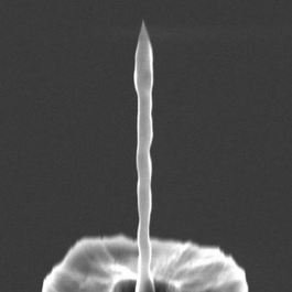
| Tip shape | Colloidal ball |
One of cantilevers for colloidal particle attachement can be choosen:
| Cantilever type | A | B | Typical dispersion |
| Length, L (µm) | 223 | 183 | ± 2 |
| Width, W (µm) | 34 | 34 | ± 3 |
| Thickness, H (µm) | 3 | 3 | ± 0.15 |
| Force Constant (N/m) | 3.5 | 6 | ±20% |
| Resonant frequency (kHz) | 77 | 114 | ± 10% |
| Material type | Diameters available, µm |
| SiO2 | 2.01; 3.5; 4.63; 6.62; 10; 15; 28.75 |
| PMMA | 1.5; 3.36; 6.44; 10; 108 |
| BSG | 2; 5; 10; 20 |
| Gold | 1.5 - 3; 3 - 5.5; 5.5 - 9 |
| Glass beads | 10 - 50 |
| Barium Titanate Solid Glass | 74 - 90; 90 - 106 |
| Soda Lime Glass | 100 |
| Polysterene | 1.98; 3.6; 6.1; 10; 15 |
| Glass | 70 - 110; 180 - 212 |
Specification of HA_NC colloidal AFM probes:
| Material |
Polysilicon lever
|
| Chip size | 3.6 x 1.6 x 0.4 mm |
| Reflective side | Au |
| Cantilever number | 1 rectangular |
| Tip shape | Colloidal ball |
One of cantilevers for colloidal particle attachement can be choosen:
| Cantilever type | A | B | Typical dispersion |
| Length, L (µm) | 94 | 124 | ± 2 |
| Width, W (µm) | 34 | 34 | ± 3 |
| Thickness, H (µm) | 1.85 | 1.85 | ± 0.15 |
| Force Constant (N/m) | 12 | 3.5 | ±20% |
| Resonant frequency (kHz) | 235 | 140 | ± 10% |
10.09.2018
 We're glad to present the new product line in our assortment: Etalon Premium probes with pencil-shape tips for high-quality AFM scanning!
In our experience (and by reports of our customers), these probes show better scanning data for almost all types of surfaces. They are especially effective for high objects of 1 um and higher.
At the moment Etalon Premium probes presented only with HA_NC-based lever parameters. But in the first half of 2019 we plan to expand this product line by HA_C, HA_CNC, HA_FM and HA_HR Premium models. Condutive Etalon Premium cantilevers will be also designed soon.
More detailed technical data can be found on Etalon Premium product page. We hope that our new products will suit well for your investigations!
03.09.2018
 In the end of August together with our colleagues from Ostec-Instruments we were attending International conference Scanning Probe Miscroscopy - 2018 in Ekaterinburg.
We appreciate our colleagues from Ural Federal University - for warm wellcome and excellent organization of all events and lectures, and many interesting people who we met during the conference - for great pastime and valuable ideas for our next developments and common projects.
12.06.2018
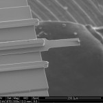
 The necessity of tip's shape estimation arises very often during AFM measurements. Even if manufacturer claims this parameter with high accuracy, fast tip's wear off during scanning leads to the need of its repeated re-calculation.
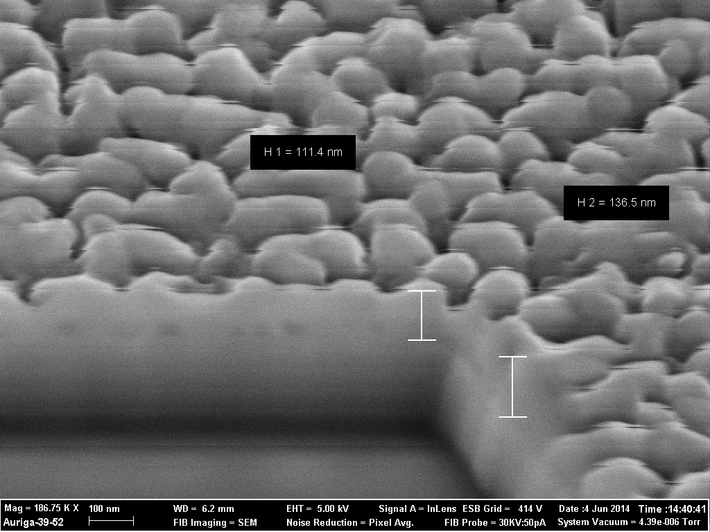
One of the most popular and cheapest methods to control tip's apex is so-called "blind" tip's shape estimation, suggested by J.S. Villarrubia in 1997. Now it is implemented in many different AFM-image-processing programs. But proper choice of a test sample for such investigation is also a tricky task. It should include many densely packed and respectively hard particles, which size will of the same order that tip's curvature radius.
Today we present our new product: TSD01, the test sample for tip's curvature radius estimation. TSD01 consists of large variety of densely packed particles with average diameter around 60 nm. Particles’ shape is not ideally round. Some of them are rather cylindrical, some ones have got vertical walls and sharp corners (like as on the REM photo). These features are necessary to collect enough statistics for further “blind” tip estimation using Deconvolution algorithm.
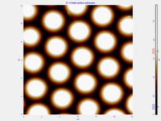
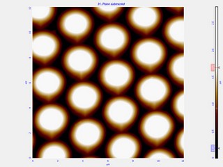
Another possible usage of this test sample - qualitative tip's quality control by simple comparison of TSD01 scans "before" and "after" scanning.
More information about TSD01 is presented on its product page. We will be also glad to discuss deconvolution method, its possible applications and correct usage of TSD01 for it. Please, contact us if any questions will appear.